AY Honor Bones, Muscles, and Movement Answer Key
Skill Level
2
Year
1999
Version
25.04.2024
Approval authority
General Conference
1
f.f An exoskeleton is a hard shell on the outside of a creature (such as an insect or a lobster). An endoskeleton is the system of bones on the inside of a creature (such as a human, dog, cat, or a bird).
2
- The skeletal system provides support to a body. Without a skeleton, a Pathfinder would be a shapeless blob.
- The marrow inside bones produces blood cells.
- The skeletal system protects the internal organs from physical harm.
- Bones serve as a place where the body can store minerals for later use.
- Bones also serve as levers against which the muscles pull to accomplish motion.
3
Bone is living tissue. Were it not so, it could not produce blood cells, nor
could bones heal after being broken. Bones cells continually regenerate
themselves.
4
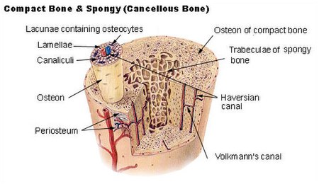
Bone is a relatively hard and lightweight composite material, formed mostly of calcium phosphate. It has relatively high compressive strength but poor tensile strength. While bone is essentially brittle, it is somewhat elastic due to its organic components (chiefly collagen). Bone has an internal mesh-like structure, the density of which may vary at different points.
Bone can also be either woven or lamellar. Woven bone is put down rapidly during growth or repair. It is so called because its fibres are aligned at random, and as a result has low strength. In contrast lamellar bone has parallel fibres and is much stronger. Woven bone is often replaced by lamellar bone as growth continues.
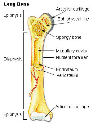
Bone growth begins with points in the cartilage. Most bones appear during fetal development, though a few short bones begin primary ossification after birth. Secondary ossification occurs after birth, and forms the ends of the long bones (the epiphyses) and the outer parts of irregular and flat bones. The main shaft (diaphyses) and the ends of long bones remain separated by a growing zone of cartilage (the metaphysis) until the child reaches skeletal maturity (18 to 25 years of age), which is when the cartilage ossifies, fusing the two together.
Marrow can be found in many of the larger bones. In newborns, all such bones are filled exclusively with red marrow, but as the child ages it is mostly replaced by yellow marrow (or fatty marrow). In adults, red marrow is mostly found in the flat bones of the skull, the ribs, the vertebrae and pelvic bones.
5
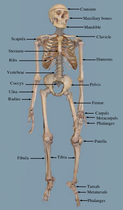
This image shows all the bones listed above. It should be noted that the maxillary bones are the bones of the face. Also note that the ulna connects to the humerus and is larger than the radius. The radius connects to the ulna. The phalanges are both the finger bones and the toe bones.
6
A joint is the place where two bones come into contact with one another.
7
Fibrous joints are where the bones are connected together by a fibrous connective tissue. These joints allow for little to no movement. An example of fibrous joints are the joints between the various bones in the cranium.
Cartilagenous joints are connected by cartilage. These joints allow for little movement. An example of a cartilagenous joint is the joint between the ribs and the sternum.
Synovial joints are separated by an empty space called a synovial cavity. These joints allow the most movement. Examples of a synovial joints are the elbow and the knee.
8
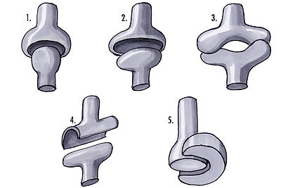
- Ball and Socket joints can be found in the hip. These joints allow for movement in many directions.
- Ellipsoid joints can be found in the knee. When the knee is extended, it does not allow rotation. When it is flexed, it allows for limited rotation.
- Saddle joints can be found in the fingers and thumbs.
- Hinge joints can be found in the elbow between the humerus and the ulna. A hinge joint works just like a door hinge, allowing motion in only one direction.
- Pivot joints can also be found in the elbow between the ulna and the radius. This type of joint allows one bone to rotate about the other.
- Gliding joints can be found between the carpals in the wrists. These joints allow limited movement between the bones as one glides past the other.
9
Approaches to constructing a model include:
- Form the bones with clay and then let them harden.
- Cover wooden dowels with papier-mâché.
- Make a wire frame and cover that with papier-mâché.
- Carve bones out of soft wood (such as balsa or pine).
- Make an impression in wet sand and fill with plaster.
- Make cross-sections out of paperboard and glue them together.
- Make the bones from sugar cookie dough and then bake them (a saddle joint or gliding joint works best for this approach).
- Use Pathfinders to model the bones and muscles in a joint. A simpler joint like the elbow is easiest. Make sure each Pathfinder knows the name and function of the body part he/she is modeling.
Be sure to review requirement 17 before starting. It's important to know how this model will be used during the design phase.
10
Another name for a broken bone is "fracture." There are two main types of fractures, open and closed. In an open fracture (also called a compound fracture), the bone protrudes through the skin. This is a very serious condition as it allows infection to enter the body. In a closed fracture (also called a simple fracture), the bone stays inside the body.
Sub-categories of fractures include:
Transverse - the break in the bone is at a right angle to the long axis of the bone.
Comminuted - the bone breaks into three or more pieces.
Greenstick - the bone breaks on one side, and the other side is bent. This is very similar to what happens when a green twig is snapped: it doesn't come all the way apart, but it is clearly broken on one side.
Stress - a hairline crack in the bone. The bone does not separate into two pieces.
Living bones are in constant change. Cells die and are replaced on a regular basis. Because of this, a bone will heal all by itself if allowed to. Doctors can help this process in two ways. First, they can set the bone. This is done by making sure the pieces of the bone are aligned properly. In a comminuted fracture this may involve surgery. The second way that doctors help bones to heal is by immobilizing them - that is, keeping them from moving around. This can be done by surrounding the broken limb with a cast, or it can be done by embedding pins inside the body. Some bones do not need to be immobilized when broken (such as the nose).
11
Osteoporosis is the depletion of minerals from the bone. Minerals in bones give them their strength, so a bone with a low bone mineral density (or BMD) is more susceptible to breakage, even when stressed only lightly. Anyone can get osteoporosis, but it is more common in postmenopausal women. Some of the risk factors of getting osteoporosis cannot be reduced by modifying behavior (such as being a woman, having dementia, having a family history of weak bones, or being of European descent), but other factors are easily avoided by healthful living. These health habits include:
- Avoid soft drinks, especially those containing phosphoric acid.
- Avoid alcohol.
- Avoid tobacco.
- Maintain a healthy body weight. Those with abnormally low body weight are more susceptible to osteoporosis.
- Make sure your diet has plenty of calcium.
- Make sure your diet has plenty of vitamin D.
- Get plenty of exercise.
12
The muscular system is the system in humans and in animals that facilitate motion. This motion can be either internal (a beating heart) or external (walking).
13
- Cardiac muscles are found only in the heart. These muscles are what make your heart beat. Cardiac muscles are the only muscles in the body consisting of branching fibers.
- Skeletal muscles are attached to the skeleton and are responsible for voluntary (and sometimes involuntary) bodily movement.
- Smooth muscles are responsible for internal, involuntary (and sometimes voluntary) movement of the organs such as breathing and moving food through the digestive tract.
14
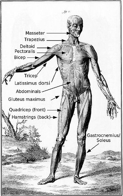
The trapezius, triceps, latissimus dorsi, gluteus maximus, hamstrings, gastocnemius, and soleus are located on the back of a human, and the arrows indicate their location.
The gluteus maximus is more commonly known as the buttock.
The gastrocnemius and soleus muscles are considered to be the same muscle by some anatomists because they share a tendon. The soleus is beneath the gastrocnemius which is closer to the skin. Together, they form the muscle more commonly known as the calf.
15
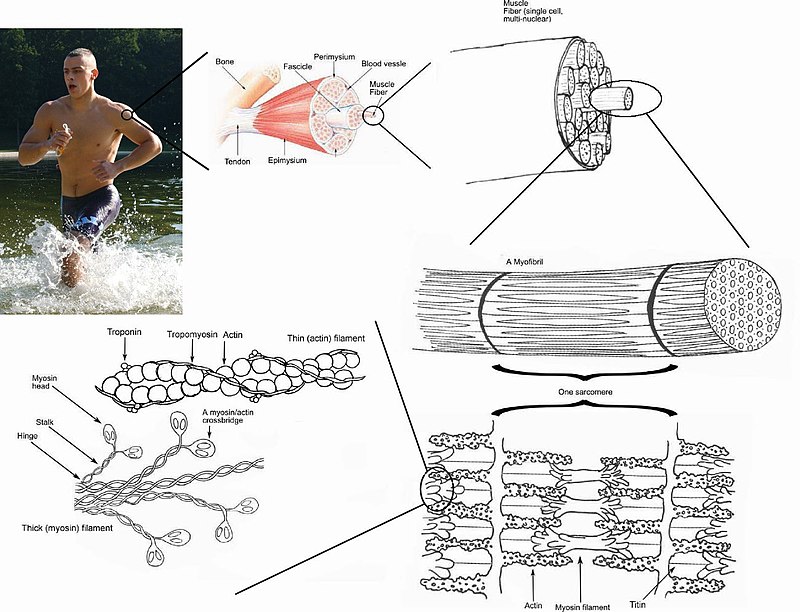
- When the muscle is in a resting state, thin strands of a protein called tropomyosin are wrapped around the actin filaments, blocking the myosin binding sites. This keeps the myosin from binding to actin.
- Molecules called troponin are attached to the tropomyosin.
- When calcium is introduced into the muscle cell, calcium ions bind to troponin molecules.
- Calcium then pulls troponin, causing tropomyosin to be moved as well, therefore causing the myosin binding sites on the actin to be exposed.
- Myosin binds to the now-exposed binding sites.
- As soon as the myosin head binds to actin, the head bends at its hinge.
- Once the head bends, the myosin loses energy, and remains attached to the actin.
- When re-energized by adenosine triphosphate (ATP), the myosin head detaches from the actin filament, and is ready to attach and bend again.
- The collective bending of numerous myosin heads (all in the same direction), combine to move the actin molecules closer together. This results in a muscle contraction.
16
Voluntary muscles are attached to the skeletal frame and are generally under the control of the person to whom they belong. These are the muscles in the arms and legs (and elsewhere). Involuntary muscles are controlled by the autonomic system without the conscious control by the individual. Your heart continues to beat, you continue to breathe, and you digest your food even when you are not thinking about it.
17
Please note that the answers given here are only possibilities. If you know a creative way to do this, you are encouraged to add it.
- Use rubberbands or elastic as muscles. Stretch them such that the joint can be moved by hand and then snap back into position when the rubber band (muscle) contracts.
- If you use cookie dough, you may have a hard time keeping the bones from breaking. You may wish to form the dough around something a little more substantive, such as a dowel, a stick of celery, or perhaps a carrot. In this case you may use Twizzlers as muscles. Although they will not contract, they look kind of like muscles and taste pretty good.
- If you use clay, plaster, or papier-mâché, be sure to make anchor points for the muscles before the "bones" harden. Try using cup hooks or eye hooks for this purpose. If using a wire frame, you can make the anchors out of the wire.
- If you use Pathfinders to model the joint, have them pull alternately (since muscles do not push) on the Pathfinders who are modeling the bones of the joint, to achieve the proper motion of the joint in all of its proper directions. This is usually more easily achieved if everyone is lying down, so gravity is less of a factor.
18
Note: In many translations of the Bible, the term "flesh" is used to describe muscles. Here are several passages featuring bones and muscles. Encourage your Pathfinders to look through more than the required three verses. It will be easier for them to describe the passage in their own words if they have made a connection with the verses.
- Genesis 2:21-24
- Exodus 12:46
- Job 40:16
- Job 10:10-12
- Psalm 22:14
- Ezekiel 37:1-12
- John 19:36
- Psalm 139:13-16
