Difference between revisions of "AY Honors/Microscopic Life/Answer Key"
m (/* 2. Be able to identify the following parts of a microscope and explain or demonstrate the function of each: eye-piece or ocular, objective, body tube, nosepiece, stage, diaphragm, base, focus knob,) |
m (- Category of AYHAB) |
||
| (53 intermediate revisions by 9 users not shown) | |||
| Line 1: | Line 1: | ||
| − | {{ | + | {{HonorSubpage}} |
| − | + | <!--{{Honor_Master|honor=Microscopic Life|master=Naturalist|group=Fauna}}--> | |
| − | ==2. Be able to identify the following parts of a microscope and explain or demonstrate the function of each: eye-piece or ocular, objective, body tube, nosepiece, stage, diaphragm, base, focus knob, and arm. | + | <!--{{Honor_Master|honor=Microscopic Life|master=Zoology|group=Fauna}}--> |
| + | <section begin="Body" /> | ||
| + | {{ansreq|page={{#titleparts:{{PAGENAME}}|2|1}}|num=1}} | ||
| + | <noinclude><translate></noinclude> | ||
| + | <!-- 1. List four major types of microscopes. What are some of the characteristics of each? Be able to identify the different types of microscopes from pictures, or visit a laboratory in a university or industry which has these microscopes. --> | ||
| + | ===Optical Microscope=== <!--T:26--> | ||
| + | This is the "classic" form of a microscope which uses optical lenses to magnify the specimen for observation. | ||
| + | |||
| + | ===Electron Microscope=== <!--T:2--> | ||
| + | The electron microscope is a type of microscope that uses electrons to create an image of the target. It has much higher magnification or resolving power than a normal light microscope, up to two million times, allowing it to see smaller objects and details. | ||
| + | ===Dark Field Microscope=== | ||
| + | Dark field microscopy is an optical microscopy illumination technique used to enhance the contrast in unstained samples. It works on the principle of illuminating the sample with light that will not be collected by the objective lens, so not form part of the image. This produces the classic appearance of a dark, almost black, background with bright objects on it. | ||
| + | ===Fluorescence Microscope=== | ||
| + | A Fluorescence Microscope is a light microscope used to study properties of organic or inorganic substances using the phenomena of fluorescence and phosphorescence instead of, or in addition to, reflection and absorption. | ||
| + | |||
| + | <!--T:3--> | ||
| + | Fluorescence is a luminescence in which the absorption of a photon triggers the release of another photon with a longer wavelength. Usually the absorbed photon is in the ultraviolet range, and the emitted light is in the visible range. | ||
| + | |||
| + | <!--T:4--> | ||
| + | Phosphorescence is a specific type of photoluminescence related to fluorescence. Unlike fluorescence, a phosphorescent material does not immediately re-emit the radiation it absorbs. The most familiar form of phosphorescence is "glow-in-the-dark" materials such as found on the hands of a watch. | ||
| + | |||
| + | ===Phase Contrast=== <!--T:5--> | ||
| + | A phase contrast microscope is a microscope that does not require staining to view the slide. This microscope made it possible to study the cell cycle. | ||
| + | |||
| + | <!--T:6--> | ||
| + | As light travels through a medium other than vacuum, interaction with this medium causes its amplitude and phase to change in a way which depends on properties of the medium. Changes in amplitude give rise to familiar absorption of light which gives rise to colours when it is wavelength dependent. The human eye measures only the energy of light arriving on the retina, so changes in phase are not easily observed, yet often these changes in phase carry a large amount of information. | ||
| + | |||
| + | <!--T:7--> | ||
| + | The same holds in a typical microscope, i.e., although the phase variations introduced by the sample are preserved by the instrument (at least in the limit of the perfect imaging instrument) this information is lost in the process which measures the light. In order to make phase variations observable, it is necessary to combine the light passing through the sample with a reference so that the resulting interference reveals the phase structure of the sample. | ||
| + | |||
| + | <!--T:27--> | ||
| + | <noinclude></translate></noinclude> | ||
| + | {{CloseReq}} <!-- 1 --> | ||
| + | {{ansreq|page={{#titleparts:{{PAGENAME}}|2|1}}|num=2}} | ||
| + | <noinclude><translate><!--T:28--> | ||
| + | </noinclude> | ||
| + | <!-- 2. Be able to identify the following parts of a microscope and explain or demonstrate the function of each: eye-piece or ocular, objective, body tube, nosepiece, stage, diaphragm, base, focus knob, and arm. --> | ||
[[Image:Mikroskop.png|frame| | [[Image:Mikroskop.png|frame| | ||
1. eye-piece or ocular<br> | 1. eye-piece or ocular<br> | ||
| Line 12: | Line 48: | ||
8. diaphragm and condenser | 8. diaphragm and condenser | ||
]] | ]] | ||
| − | ;The eyepiece: The part of a microscope that a user looks into. | + | ;The eyepiece or ocular (1): The part of a microscope that a user looks into. It contains a lens called the ''ocular''. |
| − | ;Objective | + | ;Objective (3): The objective is another lens. It is located near the specimen to be observed. |
| − | ;Body Tube: | + | ;Body Tube: This is a hollow tube that connects the ocular lens to the objective lens. |
| − | ;Nosepiece | + | ;Nosepiece (2): The nosepiece is the part of the microscope that the objective lenses attach to. Sometimes the nosepiece houses a prism whose function is to bend light from the image so that the user can sit comfortably instead of hunching over the microscope to look straight down on the specimen. |
| − | ;Stage | + | ;Stage (6): The stage is a platform where the slides are mounted. |
| − | ; | + | ;Diaphragm (8): The diaphragm is an apparatus located beneath the stage. It focuses light onto the specimen. |
| − | ;Base: | + | ;Base: The base is the bottom of the microscope on which the rest of the instrument rests. |
| − | ;Focus knob | + | ;Focus knob (4 and 5): The focus knob (or knobs) adjust the distance between the ocular lens and the objective lens. This brings the specimen into focus. Microscopes often come with two focus knobs - a coarse focus and a fine focus. The coarse focus knob makes large changes in the focus. The fine focus know makes smaller adjustments. |
| − | ;Arm: | + | ;Arm: The arm connects to the base and other parts of the microscope (such as the stage, diaphragm, and body tube) attach to it. |
{{TODO|edit the picture and renumber so the parts we need are in the order presented here}} | {{TODO|edit the picture and renumber so the parts we need are in the order presented here}} | ||
| − | ==3. Know how to calculate the magnification of a compound microscope. Calculate the magnification of the microscope you use for this honor.== | + | <!--T:9--> |
| − | ==4. Define the following microscopic terms: slide, coverslip, wetmount, fixing, staining, oil immersion, unicellular, multicellular, cilia, flagella, plankton. | + | <br style="clear:both"> |
| − | ;slide: | + | |
| − | ;coverslip: | + | <!--T:29--> |
| − | ;wetmount: | + | <noinclude></translate></noinclude> |
| − | ;fixing: | + | {{CloseReq}} <!-- 2 --> |
| − | ;staining: | + | {{ansreq|page={{#titleparts:{{PAGENAME}}|2|1}}|num=3}} |
| − | ;oil immersion: | + | <noinclude><translate><!--T:30--> |
| − | ;unicellular: | + | </noinclude> |
| − | ;multicellular: | + | <!-- 3. Know how to calculate the magnification of a compound microscope. Calculate the magnification of the microscope you use for this honor. --> |
| − | ;cilia: | + | The magnification of a compound microscope is simply the magnification of the ocular lens times the magnification of the objective lens: |
| − | ;flagella: | + | |
| − | ;plankton: | + | <!--T:11--> |
| + | <math>magnification = ocular \times objective</math> | ||
| + | |||
| + | <!--T:12--> | ||
| + | Therefore, if a microscope's objective lens has a magnification of 10 and its objective lens has a magnification of 40, the microscope's magnification is | ||
| + | |||
| + | <!--T:13--> | ||
| + | <math> 10 \times 40 = 400X</math> | ||
| + | |||
| + | <!--T:31--> | ||
| + | <noinclude></translate></noinclude> | ||
| + | {{CloseReq}} <!-- 3 --> | ||
| + | {{ansreq|page={{#titleparts:{{PAGENAME}}|2|1}}|num=4}} | ||
| + | <noinclude><translate><!--T:32--> | ||
| + | </noinclude> | ||
| + | <!-- 4. Define the following microscopic terms: slide, coverslip, wetmount, fixing, staining, oil immersion, unicellular, multicellular, cilia, flagella, plankton. --> | ||
| + | ;slide: A slide is a small piece of rectangular glass upon which the specimen to be viewed is placed. | ||
| + | ;coverslip: The coverslip is a piece of glass the same shape as a slide (but often thinner) used to cover the specimen. The specimen is sandwiched between the slide and the coverslip. | ||
| + | ;wetmount: Wetmounting is when the user smears a wet specimen onto a slide. | ||
| + | ;fixing: Fixing preserves a specimen so that it does not decompose. Once a specimen has been fixed, it can be stored away and looked at again later. | ||
| + | ;staining: Staining colors the specimen so that it has a higher contrast and can be more easily seen under the microscope. | ||
| + | ;oil immersion: In order to get a sharp focus at magnifications above 400X, light must be coupled between the specimen and the objective by a layer of oil. If the light travels through air it gets too distorted. | ||
| + | ;unicellular: A unicellular organism has only one cell. This includes bacteria, many algae, amoebae, and many other organisms that cannot be seen with the naked eye. | ||
| + | ;multicellular: A multicellular organism is made up of more than one cell. This includes animals, insects, plants, and most other life that can be seen around us on a daily basis. | ||
| + | ;cilia: Cilia are small hair-like appendages around the edge of a cell which allows the cell to propel itself through water. Cells in the human body have cilia. The cilia in the respiratory tract move mucus with particles trapped out of the air we breathe back up into the throat. | ||
| + | ;flagella: A flagella is a whip-like structure at the end of a cell that allows it to swim through the water. This is the way that sperm swim. Many bacteria also have flagella for swimming through their environment. | ||
| + | ;plankton: Plankton are any type unicellular marine organism at the bottom of the food chain. | ||
| + | |||
| + | <!--T:33--> | ||
| + | <noinclude></translate></noinclude> | ||
| + | {{CloseReq}} <!-- 4 --> | ||
| + | {{ansreq|page={{#titleparts:{{PAGENAME}}|2|1}}|num=5}} | ||
| + | <noinclude><translate><!--T:34--> | ||
| + | </noinclude> | ||
| + | <!-- 5. Collect samples of water (from ponds, streams, ditches, gutters, puddles, etc.) And search for living organisms using a microscope with at least 100X magnification. Draw five of these organisms as accurately as possible. As far as possible, identify and label your diagrams (include the magnification used.) --> | ||
| + | You will have better luck with this in the summer than in the winter, though it is not difficult to find microscopic life even in the winter. Still water is more likely to harbor microscope life than swift water. Often scraping 'scum' off of rocks will yield an interesting specimen. If necessary, instruct your Pathfinders to harvest a piece of ice from a frozen puddle in the woods or in a ditch, and let it thaw out before coming to the meeting. Aquariums and flower vases are good sources of water laden with microscopic life. Another option is to seed some tap water and let it "marinate" for a week. You can seed the water with hay, straw, grass, or even dirt from the floor. Just don't get ''too'' gross! | ||
| + | |||
| + | <!--T:35--> | ||
| + | There are '''so''' many forms of microscopic life that it is highly unlikely that you will be able to identify most of what you see under the microscope. If you cannot identify what you see, draw it anyhow, and label the parts of the cell you can identify (such as the nucleus, cell membrane, and cytoplasm). | ||
| + | |||
| + | <!--T:16--> | ||
| + | An additional way to observe cells in multicellular life is to make an impression of plant leaves. Paint the underside of a leaf with a thin layer of clear nail polish. After it has dried, carefully peel off the thin layer and place it on a slide. Using a coverslip will flatten the nail polish layer and keep the entire viewing plane in focus. This technique allows observation of detailed impressions of stomata (the opening that allow for gas exchange in plant leaves) and the guard cells that surround the stomata opening. Detailed directions can be found at: http://www.microbehunter.com/using-clear-nail-polish-to-make-impressions-of-plant-leaves/. Start out with some sturdy, live leaves. Try different colored leaves and you can often see pigments as well. | ||
| + | |||
| + | <!--T:36--> | ||
| + | <noinclude></translate></noinclude> | ||
| + | {{CloseReq}} <!-- 5 --> | ||
| + | {{ansreq|page={{#titleparts:{{PAGENAME}}|2|1}}|num=6}} | ||
| + | <noinclude><translate><!--T:37--> | ||
| + | </noinclude> | ||
| + | <!-- 6. Draw and label a cell which includes the following parts: cell membrane, nucleus, and cytoplasm. --> | ||
| + | [[Image:Cell parts.png|thumb|400px|center|Basic parts of a cell]] | ||
| + | <br style="clear:both"> | ||
| + | |||
| + | <!--T:38--> | ||
| + | <noinclude></translate></noinclude> | ||
| + | {{CloseReq}} <!-- 6 --> | ||
| + | {{ansreq|page={{#titleparts:{{PAGENAME}}|2|1}}|num=7}} | ||
| + | <noinclude><translate><!--T:39--> | ||
| + | </noinclude> | ||
| + | <!-- 7. Know the kingdoms that have microscopic life forms and know two members from each. --> | ||
| + | There are six kingdoms into which living organisms are divided. All of them contain microscopic forms of life, and some of them are composed exclusively of microscopic life. Prior to 1977, Eubacteria and Archaeabacteria were considered a single kingdom called ''Monera''. | ||
| + | |||
| + | <!--T:19--> | ||
| + | ;Animalia: Roundworms, flatworms, tapeworms, nematodes, flukes | ||
| + | ;Plantia: plant spores, green algaes | ||
| + | ;Fungi: Yeast, Penicillin, ringworms | ||
| + | ;Protista: Algae, Euglena, Amoeba, Paramecium, Toxoplasma | ||
| + | ;Eubacteria: Streptococcus, E. Coli | ||
| + | ;Archaeabacteria: Methanopyrus, Picrophilus | ||
| + | |||
| + | <!--T:40--> | ||
| + | <noinclude></translate></noinclude> | ||
| + | {{CloseReq}} <!-- 7 --> | ||
| + | {{ansreq|page={{#titleparts:{{PAGENAME}}|2|1}}|num=8}} | ||
| + | <noinclude><translate><!--T:41--> | ||
| + | </noinclude> | ||
| + | <!-- 8. Give at least one example of how microscopic life is important for: human food, human health, medicine, other organisms. --> | ||
| + | ;Human food: Leavened bread and cheese would not be possible without microscopic fungi. | ||
| + | ;Human health: Most bacteria is actually helpful for human health and we have many microbes on our body when we are healthy. The stomach is filled with bacteria which help break down food. The skin naturally has bacteria that crowd out unwanted bacteria. | ||
| + | |||
| + | <!--T:42--> | ||
| + | Find more resources and teaching tools on the human microbiome at: | ||
| + | http://learn.genetics.utah.edu/content/microbiome/ | ||
| + | https://www.asm.org/index.php/colloquium-reports/item/4476-humanmicrobiome | ||
| + | https://commonfund.nih.gov/hmp/public | ||
| + | |||
| + | <!--T:43--> | ||
| + | ;Medicine: There are some bacteria that cause illness, including strep, staph, salmonella, and E. coli. Viruses are another form of microscopic life that can cause illness, including the flu, the common cold, and HIV-AIDS.'''Penicillin''' is a fungi that is also a powerful antibiotic. '''Vaccination''' is the process of administering weakened or dead disease-causing microscopic lifeforms to a healthy person or animal, with the intent of conferring immunity against a targeted form of a related disease agent. Medical science could hardly even be called science before the discovery of microscopic life. It is through the microscope that we know anything about most diseases. | ||
| + | ;Other organisms: '''Lichens''' are symbiotic associations of a fungus with a photosynthetic partner that can produce food for the lichen from sunlight. Both the fungus and the photosynthetic partner start out as microscopic. Most plants would not be able to grow well at all (much less produce healthy fruits and veggies) without the bacteria and fungi that live in and around their root. Most people learn that trees are what produce the oxygen that we need to live. However, cyanobacteria in the ocean play a huge role in generating that oxygen that we breathe. | ||
| + | |||
| + | <!--T:44--> | ||
| + | <noinclude></translate></noinclude> | ||
| + | {{CloseReq}} <!-- 8 --> | ||
| + | {{ansreq|page={{#titleparts:{{PAGENAME}}|2|1}}|num=9}} | ||
| + | <noinclude><translate><!--T:45--> | ||
| + | </noinclude> | ||
| + | <!-- 9. Give at least three health habits that have been established as a direct result of harmful microscopic life. Put these habits into practice. --> | ||
| + | ;Hand-washing: Frequent hand-washing carries disease-causing germs away from the body. You should wash your hands every time you finish using the bathroom. | ||
| + | ;Tooth-brushing: Brushing your teeth checks bacteria in the mouth that cause cavities and gum disease. | ||
| + | ;Vaccinations: Vaccinations cause the body to develop immunity to deadly diseases. Smallpox was eliminated worldwide as a result of vaccination programs. Polio has been reduced to near extinction in most areas of the world due to vaccination. You can see the current status of polio eradication on the globe at http://polioeradication.org/polio-today/polio-now/. | ||
| + | ;Clean clothing: Changing into clean clothing every day - particularly the socks and underwear - can prevent sickness such as athlete's foot and jock itch. | ||
| + | ;Refrigeration: Keeping food cold can slow the overgrowth of microorganisms to levels that are harmful to the human body. | ||
| + | ;Cleaning: Removing scum from showers and other moist environments, keeping counters and other surfaces free of crumbs and dust, wiping off inanimate objects (door handles, gym equipment, etc), and all the other myriad ways that we keep our living and working environments clean. This keeps harmful (or dangerously high levels of) microorganisms from entering our bodies through the air, cuts/scrapes, or through our mouth/nose/eyes/ears. | ||
| − | + | <!--T:46--> | |
| − | + | <noinclude></translate></noinclude> | |
| − | + | {{CloseReq}} <!-- 9 --> | |
| − | + | <noinclude><translate></noinclude> | |
| − | + | ==References== <!--T:22--> | |
| − | ==References== | ||
| − | + | <!--T:23--> | |
| + | Text for these answers was heavily borrowed from Wikipedia. | ||
| + | <noinclude></translate></noinclude> | ||
| + | {{CloseHonorPage}} | ||
Latest revision as of 16:06, 14 July 2022
1
Optical Microscope
This is the "classic" form of a microscope which uses optical lenses to magnify the specimen for observation.
Electron Microscope
The electron microscope is a type of microscope that uses electrons to create an image of the target. It has much higher magnification or resolving power than a normal light microscope, up to two million times, allowing it to see smaller objects and details.
Dark Field Microscope
Dark field microscopy is an optical microscopy illumination technique used to enhance the contrast in unstained samples. It works on the principle of illuminating the sample with light that will not be collected by the objective lens, so not form part of the image. This produces the classic appearance of a dark, almost black, background with bright objects on it.
Fluorescence Microscope
A Fluorescence Microscope is a light microscope used to study properties of organic or inorganic substances using the phenomena of fluorescence and phosphorescence instead of, or in addition to, reflection and absorption.
Fluorescence is a luminescence in which the absorption of a photon triggers the release of another photon with a longer wavelength. Usually the absorbed photon is in the ultraviolet range, and the emitted light is in the visible range.
Phosphorescence is a specific type of photoluminescence related to fluorescence. Unlike fluorescence, a phosphorescent material does not immediately re-emit the radiation it absorbs. The most familiar form of phosphorescence is "glow-in-the-dark" materials such as found on the hands of a watch.
Phase Contrast
A phase contrast microscope is a microscope that does not require staining to view the slide. This microscope made it possible to study the cell cycle.
As light travels through a medium other than vacuum, interaction with this medium causes its amplitude and phase to change in a way which depends on properties of the medium. Changes in amplitude give rise to familiar absorption of light which gives rise to colours when it is wavelength dependent. The human eye measures only the energy of light arriving on the retina, so changes in phase are not easily observed, yet often these changes in phase carry a large amount of information.
The same holds in a typical microscope, i.e., although the phase variations introduced by the sample are preserved by the instrument (at least in the limit of the perfect imaging instrument) this information is lost in the process which measures the light. In order to make phase variations observable, it is necessary to combine the light passing through the sample with a reference so that the resulting interference reveals the phase structure of the sample.
2
- The eyepiece or ocular (1)
- The part of a microscope that a user looks into. It contains a lens called the ocular.
- Objective (3)
- The objective is another lens. It is located near the specimen to be observed.
- Body Tube
- This is a hollow tube that connects the ocular lens to the objective lens.
- Nosepiece (2)
- The nosepiece is the part of the microscope that the objective lenses attach to. Sometimes the nosepiece houses a prism whose function is to bend light from the image so that the user can sit comfortably instead of hunching over the microscope to look straight down on the specimen.
- Stage (6)
- The stage is a platform where the slides are mounted.
- Diaphragm (8)
- The diaphragm is an apparatus located beneath the stage. It focuses light onto the specimen.
- Base
- The base is the bottom of the microscope on which the rest of the instrument rests.
- Focus knob (4 and 5)
- The focus knob (or knobs) adjust the distance between the ocular lens and the objective lens. This brings the specimen into focus. Microscopes often come with two focus knobs - a coarse focus and a fine focus. The coarse focus knob makes large changes in the focus. The fine focus know makes smaller adjustments.
- Arm
- The arm connects to the base and other parts of the microscope (such as the stage, diaphragm, and body tube) attach to it.
3
The magnification of a compound microscope is simply the magnification of the ocular lens times the magnification of the objective lens:
[math]\displaystyle{ magnification = ocular \times objective }[/math]
Therefore, if a microscope's objective lens has a magnification of 10 and its objective lens has a magnification of 40, the microscope's magnification is
[math]\displaystyle{ 10 \times 40 = 400X }[/math]
4
- slide
- A slide is a small piece of rectangular glass upon which the specimen to be viewed is placed.
- coverslip
- The coverslip is a piece of glass the same shape as a slide (but often thinner) used to cover the specimen. The specimen is sandwiched between the slide and the coverslip.
- wetmount
- Wetmounting is when the user smears a wet specimen onto a slide.
- fixing
- Fixing preserves a specimen so that it does not decompose. Once a specimen has been fixed, it can be stored away and looked at again later.
- staining
- Staining colors the specimen so that it has a higher contrast and can be more easily seen under the microscope.
- oil immersion
- In order to get a sharp focus at magnifications above 400X, light must be coupled between the specimen and the objective by a layer of oil. If the light travels through air it gets too distorted.
- unicellular
- A unicellular organism has only one cell. This includes bacteria, many algae, amoebae, and many other organisms that cannot be seen with the naked eye.
- multicellular
- A multicellular organism is made up of more than one cell. This includes animals, insects, plants, and most other life that can be seen around us on a daily basis.
- cilia
- Cilia are small hair-like appendages around the edge of a cell which allows the cell to propel itself through water. Cells in the human body have cilia. The cilia in the respiratory tract move mucus with particles trapped out of the air we breathe back up into the throat.
- flagella
- A flagella is a whip-like structure at the end of a cell that allows it to swim through the water. This is the way that sperm swim. Many bacteria also have flagella for swimming through their environment.
- plankton
- Plankton are any type unicellular marine organism at the bottom of the food chain.
5
You will have better luck with this in the summer than in the winter, though it is not difficult to find microscopic life even in the winter. Still water is more likely to harbor microscope life than swift water. Often scraping 'scum' off of rocks will yield an interesting specimen. If necessary, instruct your Pathfinders to harvest a piece of ice from a frozen puddle in the woods or in a ditch, and let it thaw out before coming to the meeting. Aquariums and flower vases are good sources of water laden with microscopic life. Another option is to seed some tap water and let it "marinate" for a week. You can seed the water with hay, straw, grass, or even dirt from the floor. Just don't get too gross!
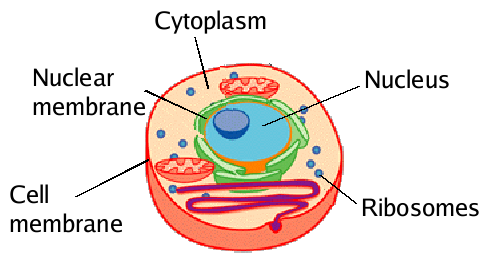
There are so many forms of microscopic life that it is highly unlikely that you will be able to identify most of what you see under the microscope. If you cannot identify what you see, draw it anyhow, and label the parts of the cell you can identify (such as the nucleus, cell membrane, and cytoplasm).
An additional way to observe cells in multicellular life is to make an impression of plant leaves. Paint the underside of a leaf with a thin layer of clear nail polish. After it has dried, carefully peel off the thin layer and place it on a slide. Using a coverslip will flatten the nail polish layer and keep the entire viewing plane in focus. This technique allows observation of detailed impressions of stomata (the opening that allow for gas exchange in plant leaves) and the guard cells that surround the stomata opening. Detailed directions can be found at: http://www.microbehunter.com/using-clear-nail-polish-to-make-impressions-of-plant-leaves/. Start out with some sturdy, live leaves. Try different colored leaves and you can often see pigments as well.
6
7
There are six kingdoms into which living organisms are divided. All of them contain microscopic forms of life, and some of them are composed exclusively of microscopic life. Prior to 1977, Eubacteria and Archaeabacteria were considered a single kingdom called Monera.
- Animalia
- Roundworms, flatworms, tapeworms, nematodes, flukes
- Plantia
- plant spores, green algaes
- Fungi
- Yeast, Penicillin, ringworms
- Protista
- Algae, Euglena, Amoeba, Paramecium, Toxoplasma
- Eubacteria
- Streptococcus, E. Coli
- Archaeabacteria
- Methanopyrus, Picrophilus
8
- Human food
- Leavened bread and cheese would not be possible without microscopic fungi.
- Human health
- Most bacteria is actually helpful for human health and we have many microbes on our body when we are healthy. The stomach is filled with bacteria which help break down food. The skin naturally has bacteria that crowd out unwanted bacteria.
Find more resources and teaching tools on the human microbiome at: http://learn.genetics.utah.edu/content/microbiome/ https://www.asm.org/index.php/colloquium-reports/item/4476-humanmicrobiome https://commonfund.nih.gov/hmp/public
- Medicine
- There are some bacteria that cause illness, including strep, staph, salmonella, and E. coli. Viruses are another form of microscopic life that can cause illness, including the flu, the common cold, and HIV-AIDS.Penicillin is a fungi that is also a powerful antibiotic. Vaccination is the process of administering weakened or dead disease-causing microscopic lifeforms to a healthy person or animal, with the intent of conferring immunity against a targeted form of a related disease agent. Medical science could hardly even be called science before the discovery of microscopic life. It is through the microscope that we know anything about most diseases.
- Other organisms
- Lichens are symbiotic associations of a fungus with a photosynthetic partner that can produce food for the lichen from sunlight. Both the fungus and the photosynthetic partner start out as microscopic. Most plants would not be able to grow well at all (much less produce healthy fruits and veggies) without the bacteria and fungi that live in and around their root. Most people learn that trees are what produce the oxygen that we need to live. However, cyanobacteria in the ocean play a huge role in generating that oxygen that we breathe.
9
- Hand-washing
- Frequent hand-washing carries disease-causing germs away from the body. You should wash your hands every time you finish using the bathroom.
- Tooth-brushing
- Brushing your teeth checks bacteria in the mouth that cause cavities and gum disease.
- Vaccinations
- Vaccinations cause the body to develop immunity to deadly diseases. Smallpox was eliminated worldwide as a result of vaccination programs. Polio has been reduced to near extinction in most areas of the world due to vaccination. You can see the current status of polio eradication on the globe at http://polioeradication.org/polio-today/polio-now/.
- Clean clothing
- Changing into clean clothing every day - particularly the socks and underwear - can prevent sickness such as athlete's foot and jock itch.
- Refrigeration
- Keeping food cold can slow the overgrowth of microorganisms to levels that are harmful to the human body.
- Cleaning
- Removing scum from showers and other moist environments, keeping counters and other surfaces free of crumbs and dust, wiping off inanimate objects (door handles, gym equipment, etc), and all the other myriad ways that we keep our living and working environments clean. This keeps harmful (or dangerously high levels of) microorganisms from entering our bodies through the air, cuts/scrapes, or through our mouth/nose/eyes/ears.
References
Text for these answers was heavily borrowed from Wikipedia.


