AY Honors/Microscopic Life/Answer Key
1. List four major types of microscopics. What are some of the characteristics of each? Be able to identify the different types of microscopes from pictures, or visit a laboratory in a university or industry which has these microscopes.
Optical Microscope
This is the "classic" form of a microscope which uses optical lenses to magify the specimen for observation.
Electron Microscope
The electron microscope is a type of microscope that uses electrons to create an image of the target. It has much higher magnification or resolving power than a normal light microscope, up to two million times, allowing it to see smaller objects and details.
Dark Field Microscope
Dark field microscopy is an optical microscopy illumination technique used to enhance the contrast in unstained samples. It works on the principle of illuminating the sample with light that will not be collected by the objective lens, so not form part of the image. This produces the classic appearance of a dark, almost black, background with bright objects on it.
Flourescent Microscope
Phase Contrast
A phase contrast microscope is a microscope that does not require staining to view the slide. This microscope made it possible to study the cell cycle.
As light travels through a medium other than vacuum, interaction with this medium causes its amplitude and phase to change in a way which depends on properties of the medium. Changes in amplitude give rise to familiar absorption of light which gives rise to colours when it is wavelength dependent. The human eye measures only the energy of light arriving on the retina, so changes in phase are not easily observed, yet often these changes in phase carry a large amount of information.
The same holds in a typical microscope, i.e., although the phase variations introduced by the sample are preserved by the instrument (at least in the limit of the perfect imaging instrument) this information is lost in the process which measures the light. In order to make phase variations observable, it is necessary to combine the light passing through the sample with a reference so that the resulting interference reveals the phase structure of the sample.
2. Be able to identify the following parts of a microscope and explain or demonstrate the function of each: eye-piece or ocular, objective, body tube, nosepiece, stage, diaphragm, base, focus knob, and arm.
- The eyepiece or ocular (1)
- The part of a microscope that a user looks into. It contains a lense called the ocular.
- Objective (3)
- The objective is another lens. It is located near the specimen to be observed.
- Body Tube
- This is a hollow tube that connects the ocular lens to the objective lens.
- Nosepiece (2)
- The nosepiece is a bend in the body tube that allows the microscope user to sit comfortably instead of hunched over the ocular looking straight down. It contains a prism that bends the light toward the viewer.
- Stage (6)
- The stage is a platform where the slides are mounted.
- Diaphram (8)
- The diaphram is an apparatus located beneath the stage. It focuses light onto the specimen.
- Base
- The base is the bottom of the microscope on which the rest of the instrument rests.
- Focus knob (4 and 5)
- The focus knob (or knobs) adjust the distance between the ocular lens and the objective lens. This brings the specimen into focus. Microscopes often come with two focus knobs - a coarse focus and a fine focus. The coarse focus knob makes large changes in the focus. The fine focus know makes smaller adjustments.
- Arm
- The arm connects to the base and other parts of the microscope (such as the stage, diaphram, and body tube) attach to it.
3. Know how to calculate the magnification of a compound microscope. Calculate the magnification of the microscope you use for this honor.
The magnification of a compound microscope is simply the magnifcation of the ocular lens times the magnification of the objective lens:
[math]\displaystyle{ magnification = ocular \times objective }[/math]
Therefore, if a microscope's objective lens has a magnification of 10 and its objective lens has a magnification of 40, the microscope's magnification is
[math]\displaystyle{ 10 \times 40 = 400X }[/math]
4. Define the following microscopic terms: slide, coverslip, wetmount, fixing, staining, oil immersion, unicellular, multicellular, cilia, flagella, plankton.
- slide
- A slide is a small piece of rectanglur glass upon which the specimen to be viewed is placed.
- coverslip
- The coverslip is a piece of glass the same shape as a slide (but often thinner) used to cover the specimen. The specimen is sandwiched between the slide and the coverslip.
- wetmount
- Wetmounting is when the user smears a wet specimen onto a slide.
- fixing
- Fixing preserves a specimen so that it does not decompose. Once a specimen has been fixed, it can be stored away and looked at again later.
- staining
- Staining colors the specimen so that it has a higher contrast and can be more easily seen under the microscope.
- oil immersion
- In order to get a sharp focus at magnifications above 400X, light must be coupled between the specimen and the objective by a layer of oil. If the light travels through air it gets too distorted.
- unicellular
- A unicellular organism has only one cell.
- multicellular
- A multicellular organism is made up of more than one cell.
- cilia
- Cilia are small hair-like appendages around the edge of a cell which allows the cell to propel itself through water.
- flagella
- A flagella is s whip-like structure at the end of a cell that allows it to swim through the water.
- plankton
- Plankton are any type unicellular marine organism at the bottom of the food chain.
5. Collect samples of water (from ponds, streams, ditches, gutters, puddles, etc.) And search for living organisms using a microscope with at least 100X magnification. Draw five of these organisms as accurately as possible. As far as possible, identify and label your diagrams (include the magnification used.)
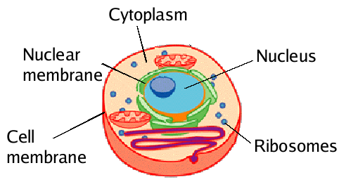
6. Draw and label a cell which includes the following parts: cell membrane, nucleus, and cytoplasm.


